
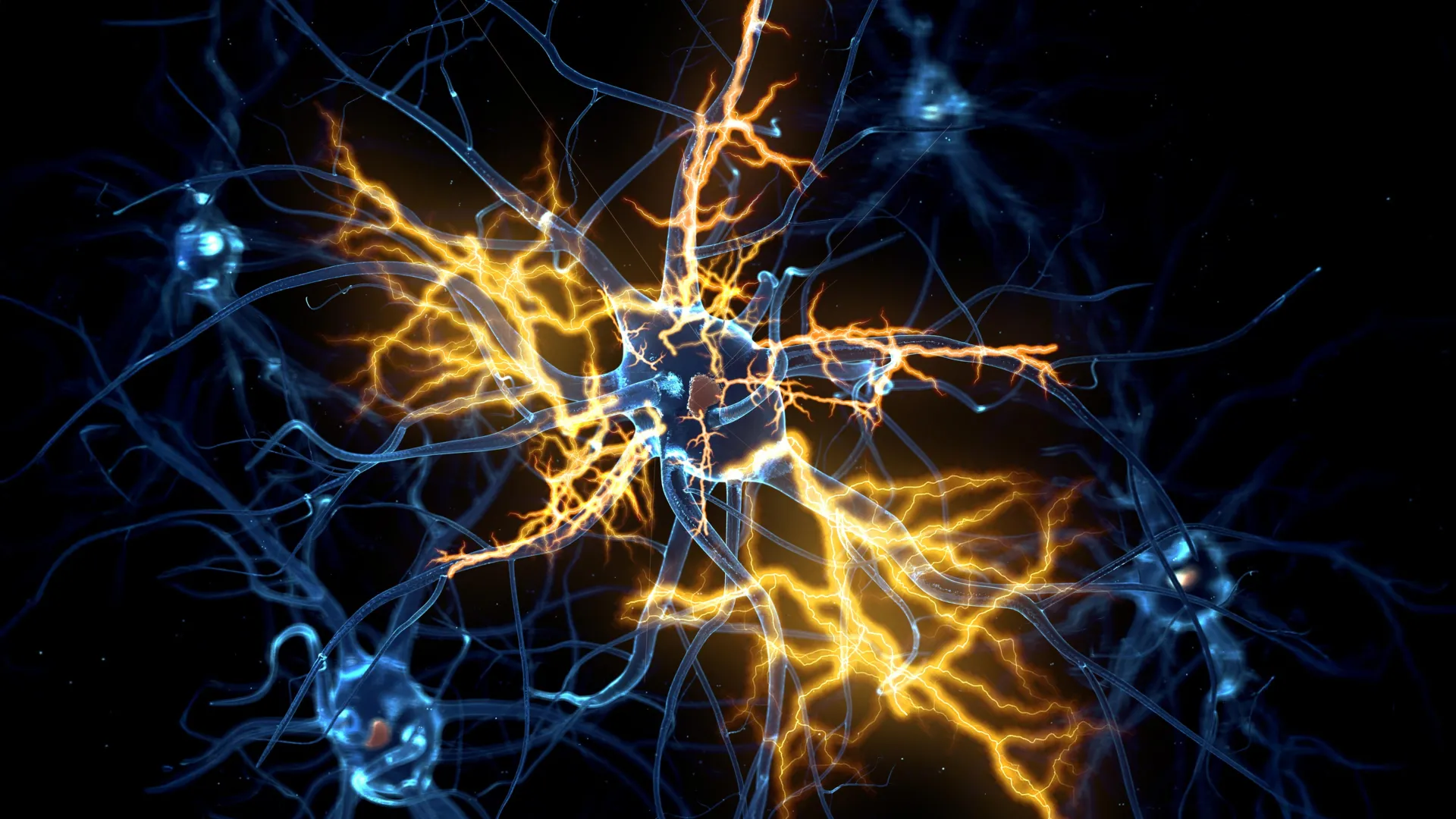
Scientists at the University of Cambridge have created tiny lab-grown brain and spinal cord systems that mimic how movement signals travel through the human nervous system. Using this model, the team discovered that nerve damage once believed to be permanent may actually be reversible under certain conditions.
As the human body develops from an embryo into a fetus and eventually an infant, neurons form complex communication networks between the brain and spinal cord. These signals travel through axons, the long nerve fibers that allow neurons to send messages and control muscle movement.
Over time, however, the central nervous system largely loses its ability to regrow damaged axons. As a result, injuries to the brain or spinal cord often become permanent, leading to serious disabilities such as paralysis or loss of movement. This loss of regenerative ability is also linked to neurological diseases including motor neurone disease and multiple sclerosis.
Mini Human Brain and Spinal Cord Models
In 2021, Dr. András Lakatos and his colleagues at the University of Cambridge developed miniature human brain models using stem cells taken from patients. These pea-sized “brain organoids” resembled parts of the cerebral cortex and allowed researchers to study molecular changes linked to motor neurone disease and explore ways to prevent them.
Now, in a new study published in Cell Reports, the researchers expanded on that work by building a miniature version of the connected human brain and spinal cord system.
Because the brain and spinal cord are separate but connected structures in the body, the team kept the organoids physically apart in the lab. They then observed axons from the brain tissue growing across the gap and connecting with the spinal cord tissue. The resulting neural circuit was functional enough to trigger contractions in tiny clusters of muscle cells.
Nerve Regrowth Declines During Development
The scientists maintained these miniature systems in the lab for more than a year. They discovered that until about day 150 of development, roughly corresponding to the middle stage of pregnancy, damaged axons could still regrow. After that point, the neurons showed a major decline in their ability to regenerate.
George Gibbons from the Department of Clinical Neurosciences at the University of Cambridge and first author of the study said: “Neurons taken from less mature organoids regrew long fibers after injury, but those from more mature organoids showed a sharp drop in their ability to regrow. In other words, poor regeneration is built into human neurons as they mature in the central nervous system.”
The team analyzed gene activity in neurons that connect the brain and spinal cord. Their work revealed a network of genes that acts like a biological switch, limiting axon growth as neurons mature and form synapses.
Remarkably, when researchers blocked key regulators within this network, the neurons regained the ability to grow axons again.
Existing Drug Boosted Nerve Regrowth
The researchers also searched a database of drug compounds to identify medicines that affect this newly identified gene network. One promising candidate was lynestrenol, a hormone drug currently approved for certain menstrual disorders and contraceptive use.
When the drug was tested on damaged neurons, it significantly improved axon regrowth.
The scientists noted that scar tissue and inflammation can also interfere with nerve repair after injury. However, understanding the neuron-specific biological mechanisms that limit regeneration remains critically important. Previous evidence has shown that younger neurons can grow through environments that normally block repair at injury sites.
Senior author Dr. András Lakatos, who led the study at the Department of Clinical Neurosciences, said: “When the brain and spinal cord are damaged, the nerve fibers that carry movement signals from the brain to the spinal cord rarely grow back. That’s why paralysis is usually permanent. But we didn’t know exactly when the ability of axons to regenerate becomes limited. Our model provides a good indication that this block happens during development, and it can still be reversed after this point.
“Lynestrenol itself may not be the answer to spinal cord repair, but it shows us that, in principle, it should be possible to directly target human neurons and regenerate their axons. Although we still need to show that this strategy will also help to re-establish appropriate connections between the brain and spinal cord cells, this gives us hope that one day we may be able to treat conditions previously thought untreatable.”
Why Human Organoids Matter
Organoid technology is becoming increasingly valuable for studying human biology and disease. While animal models such as mice and rats remain useful in research, important biological differences limit how accurately they reflect human nervous system function.
Human stem cell-derived organoids can more closely reproduce human biology, helping bridge the gap between animal experiments and real patient outcomes.
Dr. Lakatos added: “Much of what we know about nerve regeneration comes from rodents, whose neurons behave differently from human neurons. Our sophisticated organoid models help bridge the knowledge gap from animal models to what we see in patients. They are also an important contribution to efforts to reduce the use of animals in research.”
Researchers at the University of Cambridge are already using organoids for a wide range of medical studies, including efforts to repair damaged livers, investigate Crohn’s disease in children, and study the earliest stages of pregnancy.
The research was funded by the UK Research and Innovation Medical Research Council and Spinal Research.